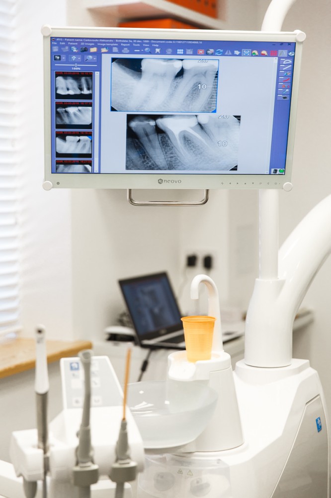
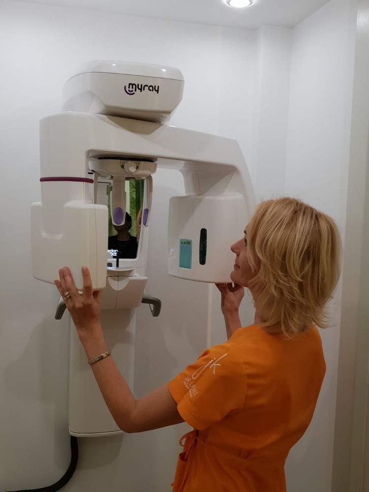
At the JK Dent dental clinic, MyRay digital X-ray equipment works for the benefit of patients.
The image is obtained immediately after the procedure. It is easy to store and available as a file, and can also be used in specialized computer modeling programs.
MyRay X-ray machines are made in Italy. All six radiographers meet the strict standards of medical equipment and use the latest X-ray imaging technologies. For doctors, they are able to provide the highest resolution images necessary for the most accurate work. It provides patients with 40% less exposure to X-rays than conventional X-ray film production.
Today’s X-rays are so gentle that there is absolutely nothing to be afraid of – X-rays are fantastically fast and produce microscopic radiation. Besides, even the best dentist can only see 50% of the tooth during the examination – the other 50% is covered by the gums and bone.
A dental X-ray is required:
- caries diagnosis and treatment;
- in the treatment of dental root canals;
- in surgery;
- in dental prosthetics
- in implantation;
In X-rays, you can get a separate picture of the teeth and you can also see what the mutual position of the teeth is, assess the bone condition, and also find out the result of previous dental procedures and, if necessary, correct it.

3-dimensional cone beam computed tomography
A 3-dimensional cone beam computer tomograph has been installed in the clinic. It can be used to perform 2D orthopantomograms, as well as 3D face and jaw examinations, which are indispensable for complex tooth extractions, endodontics and implantation. Examinations are performed with the latest generation Hyperion X5 x-ray equipment.
Objectives of maxillofacial 3D computed tomography:
- Assess the ratio of rare teeth to other teeth and their position in the bone;
- Assess the condition of endodontically treated teeth (also during treatment);
- Assess the quality and quantity of bone before placing implants;
- Assess the condition of existing implants;
- Evaluate pathological formations in the bone.

